How quickly can you have a private lumpectomy in London?
Private lumpectomy in London may reduce administrative gaps, yet no universal consultation-to-surgery interval applies. Your diagnosis, scans, pathology report and operation plan need review before a date is set. Further assessment, localisation, pre-operative checks, theatre space and anaesthetic arrangements may all affect timing.
A vacant theatre list does not set the right date on its own. The earliest clinically appropriate date follows a clear review of the information that shows where the cancer is, what surgery you need and whether any linked procedure requires planning.
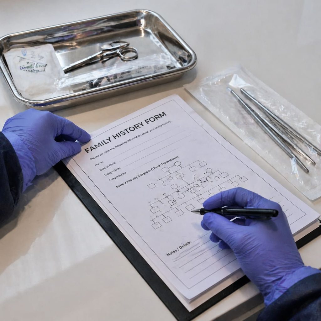
You can seek private care after an NHS diagnosis. Existing mammograms, ultrasound scans and biopsy results may provide the information needed for planning. A further scan or biopsy may be appropriate where the records leave an important question unanswered.
A maximum of 31 days applies under NHS England monitoring, from a decision to treat, or the earliest clinically appropriate date, to cancer treatment. This is an NHS monitoring standard. It does not set a private lumpectomy waiting time.
The following comparison shows what may already be available and what can still need arranging.
| Information already available | Arrangements before surgery |
|---|---|
| Mammograms or ultrasound scans that show the abnormal area | A review of whether the images show the full extent of the area |
| A biopsy report that confirms the diagnosis | Pathology review where the report needs clarification for surgery |
| A proposed operation from your existing team | Pre-operative assessment, anaesthetic planning and theatre arrangements |
| A breast abnormality that can be felt on examination | Localisation planning when the area cannot be felt |
| Insurance cover for consultation | Authorisation for tests, hospital admission or additional procedures where required |
Mr Ghosh personally reviews your diagnosis, imaging and pathology and leads your surgical planning. The clinical review determines the appropriate date, while an empty theatre slot does not.
Clinical readiness sets the surgery date
A sound operation plan starts with evidence that tells one clear clinical story. Your examination, imaging and biopsy findings guide the amount of breast tissue that needs removal and the checks needed before surgery.
The sequence commonly includes:
- Records review. Your scans and biopsy report are assessed alongside the details of your diagnosis and any previous treatment advice.
- Examination and imaging review. The surgeon checks the position and extent of the abnormal area, including its relationship to the rest of the breast.
- Pathology confirmation. A biopsy shows the type of breast cancer or whether you have ductal carcinoma in situ, which means abnormal cells contained within the breast ducts.
- Further assessment follows when the available information leaves uncertainty about the extent of disease or the best surgical approach.
- Pre-operative assessment and booking. Your health for anaesthetic, the planned procedure and hospital arrangements are reviewed before surgery is confirmed.
Breast magnetic resonance imaging, called an MRI scan, is reserved for defined clinical reasons. NICE, which covers England, advises against routine pre-operative MRI for biopsy-proven invasive breast cancer and ductal carcinoma in situ. MRI is offered where examination, mammography and ultrasound leave the extent of invasive cancer unclear. It may also be used where breast density makes mammograms difficult to assess, or where invasive lobular cancer needs sizing before breast-conserving surgery.
Armpit assessment also informs the operation plan. Ultrasound examines the lymph nodes under your arm in invasive breast cancer. An abnormal node needs needle sampling guided by ultrasound.
A sentinel lymph node biopsy removes the first draining lymph node or nodes to check for cancer spread. This procedure may form part of surgery for invasive breast cancer when ultrasound shows no lymph node involvement or needle sampling is negative. Ductal carcinoma in situ treated with breast-conserving surgery has a different pathway because sentinel node biopsy is reserved for cases with a higher risk of invasive disease.
A short period spent completing an indicated assessment can prevent an operation that fails to match the full findings.
Expert advice on breast cancer treatment, cosmetic breast surgery, and reconstruction options in London.
Book a ConsultationThe operation plan protects breast shape
A lumpectomy is usually called a wide local excision in UK breast services. It removes the affected part of the breast with a rim of surrounding breast tissue and is usually carried out under general anaesthetic.
Wide local excision forms part of breast-conserving surgery. The incision sits where it gives access to the abnormality, and the removed tissue goes to the laboratory for examination. The size and position of the area influence the surgical approach.
The abnormality can need localisation
An abnormal area that cannot be felt may need a marker so the surgeon can identify it during surgery. Image-guided localisation may use a fine wire or a magnetic seed placed with mammography or ultrasound under local anaesthetic.
The method depends on your clinical plan and the arrangements at the hospital where surgery takes place. Your surgical team coordinates localisation before the operation date.
Margins and reshaping need advance planning
During your operation, the surgical margin is the edge of the tissue removed. Laboratory examination checks whether cancer cells reach that edge. “Tumour on ink” means cancer cells are present at the marked outer edge of the specimen.
Further surgery is offered where invasive cancer or ductal carcinoma in situ is present at the radial margin. Close margins also need an individual discussion, taking account of cancer type, other health conditions, tumour features and later treatment.
An excision from a larger area of breast tissue, or from an awkward position, may affect breast shape. Oncoplastic breast surgery combines cancer removal with reshaping of the remaining breast tissue. Therapeutic mammoplasty uses breast reduction and reshaping as part of breast-conserving surgery.
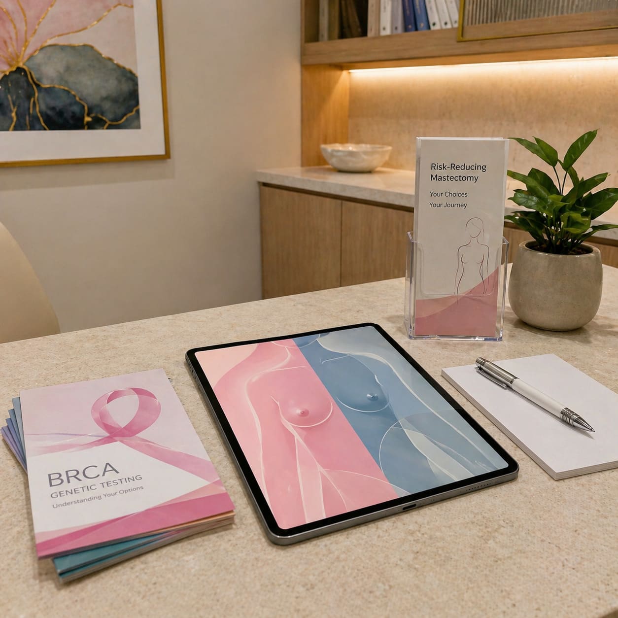
Pathology results guide any discussion about further surgery, including re-excision or mastectomy.

Before your consultation, gather your mammograms, ultrasound images, biopsy report and proposed treatment plan. This allows the surgical discussion to focus on any gaps that could affect the operation date.

Recovery and results need planning
You may go home on the day of wide local excision, although some people stay overnight. Your team confirms discharge when it is safe for you to go home after the operation.
Healing depends on the amount of tissue removed and whether your surgery includes lymph node biopsy or breast reshaping. Soreness, swelling, bruising and altered sensation may continue for several weeks. Your surgical team will give instructions on wound care, arm movement, driving and returning to normal activity.
Recognised complications include:
- Bleeding, bruising and haematoma, which is a collection of blood beneath the wound.
- Infection, pain or numbness around the breast or underarm.
- Seroma, which is a collection of fluid beneath the wound that may settle or need drainage.
- Altered breast shape, anaesthetic complications, blood clots and the possibility of further surgery.
Pathology results show what the removed tissue contains and whether the margins need further discussion. They also inform the wider treatment plan, which may include radiotherapy. Breast-conserving treatment usually includes radiotherapy, although selected people with very low-risk invasive cancer may have a different plan.
Recovery advice needs to match the operation you have had.
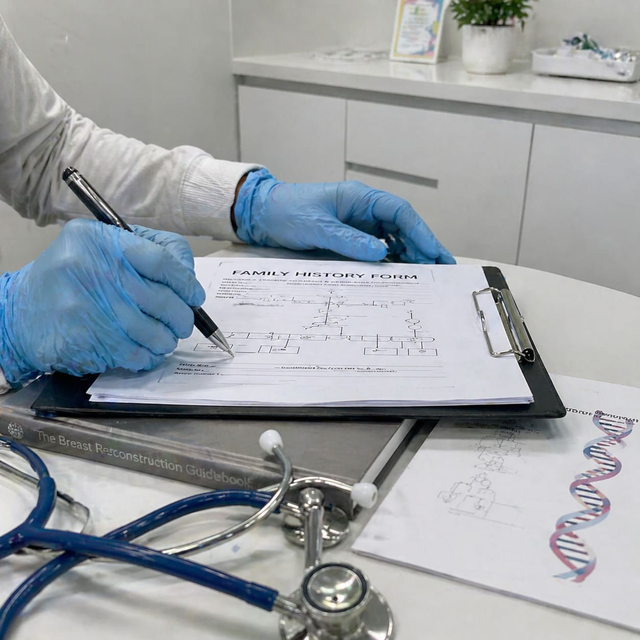
Ask how pathology results, margin findings and lymph node assessment will connect to later treatment decisions. A clear plan should explain what happens if the results lead to further surgery or radiotherapy.

Coordinated planning matters more over time
Clear handovers remain important after surgery. Imaging, pathology, surgery and later cancer treatment need to remain connected. This matters particularly when records move between NHS and private providers.
A multidisciplinary team brings together breast surgery, radiology, pathology and oncology. Their combined input helps place surgery in the right treatment sequence. A second opinion can also help where the scan findings, pathology report or proposed operation leave you uncertain.
A private pathway needs to show how external records will be reviewed, how any required localisation or lymph node assessment will fit the surgical plan, and how pathology results will feed into later treatment decisions. Consultant-led continuity keeps those decisions connected.
Expert breast surgery advice from a leading London consultant with over 30 years of experience.
Book ConsultationCommon questions about private lumpectomy
Can I have a private lumpectomy after an NHS diagnosis?
Yes. Your NHS scans, biopsy report and treatment information can be reviewed as part of private surgical planning. Further assessment may be needed where the available records do not answer an important clinical question.
Do I need an MRI before a lumpectomy?
No. MRI is used for specific clinical reasons, including unclear disease extent or difficult mammogram assessment due to breast density. Your surgeon can explain whether it adds useful information in your case.
Will I need a wire or magnetic marker before surgery?
A marker may be needed when the abnormal area cannot be felt. A fine wire or magnetic seed can help the surgeon locate the correct area during a wide local excision.
Will I need lymph node surgery with a lumpectomy?
Lymph node surgery depends on your diagnosis and armpit assessment. Sentinel lymph node biopsy may be used for invasive breast cancer, whereas ductal carcinoma in situ has different indications.
Can I drive and work soon after a lumpectomy?
Your return to driving and work depends on the surgery performed, wound healing and your comfort with normal arm movement. Follow the advice from your own surgical team before resuming these activities.
This is general information, not medical advice.
Reviewed by Mr D. B. Ghosh, Consultant Breast Surgeon, dual European Board certified (FEBS) in Breast Surgery and Surgical Oncology, and Board Certified in Cosmetic Breast Surgery through the Royal College of Surgeons scheme. GMC 4657664.