Do you need a mammogram or a breast ultrasound?
We choose the test by the clinical question, not by preference. Age, symptoms, examination findings, previous imaging, pregnancy or lactation, breast density and risk all matter. A safe route may involve mammography, breast ultrasound, both scans, or biopsy planning where tissue diagnosis is needed.

Mammogram and breast ultrasound are not rival tests
The right answer is often one scan over the other. Mammography and breast ultrasound look at the breast in different ways, and each test is useful only when it matches the problem we are trying to solve.
Screening is not the same as symptom assessment. Screening looks for early changes in people without symptoms. Symptom assessment starts with something specific, such as a breast lump, nipple change, breast pain, an unclear previous scan, or a recall after screening. Those two routes need different thinking.
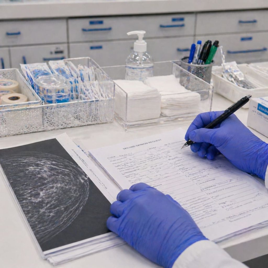
UK breast imaging guidance from The Royal College of Radiologists describes diagnostic breast assessment as triple assessment: clinical assessment, imaging and biopsy where appropriate. In plain English, we examine you, choose imaging that fits your situation, then decide whether a tissue sample is needed. That structure matters because a scan result should never be judged on its own from the breast change that brought you to clinic.
A common mistake is to treat ultrasound as the gentler replacement for a mammogram. That framing is too simple. We are asking what needs answering: a wide view of both breasts, a focused look at a lump, a check after screening recall, or a plan for biopsy if imaging and examination raise concern.
Expert advice on breast cancer treatment, cosmetic breast surgery, and reconstruction options in London.
Book a ConsultationMammography and ultrasound answer different imaging questions
A mammogram is a breast X-ray. Breast ultrasound uses high-frequency sound waves to build a picture of the inside of the breast, and it is often used to examine a focused area found by touch or on another scan.
Here is the practical difference without turning this into a radiology lesson.
| Test | What it is often used for | What it cannot decide alone |
|---|---|---|
| Mammography | A wider breast assessment, routine screening, and diagnostic assessment in the right age group | It cannot settle every felt lump without matching the result to examination and, where needed, other tests |
| Breast ultrasound | A targeted look at a lump, cyst-like change, focused pain area, or finding seen on other imaging | It does not replace mammography for routine screening and does not remove the need for biopsy where tissue diagnosis is needed |
| Both together | Checking whether the imaging picture fits the clinical finding | They still need expert interpretation, especially when symptoms and images do not line up |
Formal reports may use structured scoring, such as the British Society of Breast Radiology classification, with M1 to M5 for mammography and U1 to U5 for ultrasound. You do not need to memorise those categories. What matters is what the score means for your next decision.
A scan has value because it answers a defined question. If you can feel a lump and the scan report sounds reassuring, we still look at whether the imaging matches the exact site, size and clinical finding.
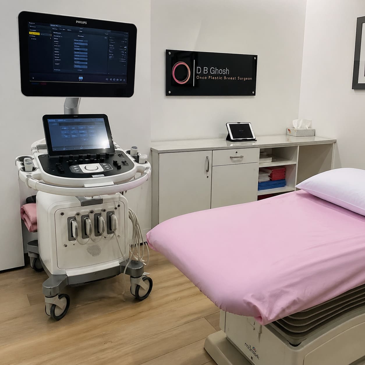
When a patient asks for a specific scan, the better question is often which clinical finding the test needs to explain. That keeps the pathway focused on diagnosis rather than preference.

Screening rules are different from symptom rules
Many people apply screening logic to a symptom-led problem, and that is where confusion starts. A recent normal screening mammogram does not mean a new breast change can wait until the next routine screen.
The NHS Breast Screening Programme offers breast screening to women aged 50 up to their 71st birthday, with first invitations between 50 and 53 and repeat invitations every 3 years until the 71st birthday. That programme is for people without symptoms. If you notice a new lump, nipple change, skin change, or another worrying breast symptom, the symptom needs assessment in its own right.
For symptomatic imaging, age affects the usual starting point. UK guidance says ultrasound is the first-line imaging test in women under 40 and during pregnancy and lactation. Mammography is the first-line test in women aged 40 or over, with ultrasound added when indicated. Those age lines guide the route, but clinical findings still matter.
A practical way to separate the routes is this:
- Routine screening: Mammography is used to look for early breast changes when you do not have symptoms. Screening does not answer every later breast concern.
- New symptom assessment: A breast lump, nipple change, skin change, or concerning new pain pattern needs clinical examination and imaging chosen around that finding. At D B Ghosh, we assess this in a London consultant-led setting so the scan choice sits inside the clinical assessment, rather than outside it.
- Age and context: Under 40, pregnancy and lactation often point first to ultrasound. From 40 onward, mammography often starts the imaging route, with ultrasound added when it helps answer the question.
Dense breast tissue can influence imaging choice, but it does not make ultrasound a simple substitute for mammography. Previous surgery, implants, family history, high-risk surveillance and unclear earlier imaging can also change the route. In those situations, the scan decision belongs inside the whole assessment, not in a separate menu of breast scan types.

Both tests may be needed to check the same finding
A felt lump is a useful example. Mammography may show broader breast patterns, while ultrasound can focus on the exact area being felt. If those findings match, the interpretation is stronger. If they do not match, the mismatch becomes part of the decision.
Combined imaging is a logic check
Needing both a mammogram and ultrasound does not automatically mean something serious has been found. Combined imaging is often used because the two tests answer different parts of the same clinical question.
A palpable lump, an abnormal screening recall, a change near previous surgery, or a finding in dense tissue may need correlation between what is felt and what is seen. We also consider whether the area needs further imaging or sampling. If invasive malignancy is suspected or confirmed, assessment may include ultrasound of the armpit area, known as axillary ultrasound, because lymph nodes can affect planning.
No imaging test should become a stand-alone promise. A normal-looking result still has to fit the examination and the history of the change.
Biopsy gives the tissue answer when imaging is not enough
Cancer Research UK says breast biopsy is the only way to find out if someone has breast cancer or another breast condition. A pathologist, a doctor who examines tissue samples, checks the sample after it is taken.
Mammography and ultrasound help decide whether biopsy is needed and where the sample should come from. A needle core biopsy takes small samples through a needle. A vacuum-assisted biopsy may be used in some situations when a larger sample is needed through a needle-based technique.
Biopsy is not automatic after ultrasound or mammography. It enters the pathway when the examination and imaging leave a question that needs tissue diagnosis. Imaging guides decisions; biopsy answers the tissue question.
A normal screening mammogram does not resolve a new symptom. A new lump, nipple change or skin change still needs diagnostic assessment against the exact site of concern.

Joined-up assessment matters more than choosing a scan
The best test is the one that changes the next decision. If a scan result does not explain your symptom, clarify the recall finding, or guide whether biopsy is needed, it has not answered the right question.
Useful clinic questions keep the assessment focused:
- What clinical question are we trying to answer with this test?
- Does the imaging match the exact lump or area I can feel?
- If the scan is normal, what explains the symptom and what follow-up is sensible?
- Would a biopsy change the plan? If so, which finding would it sample?
- How do previous surgery, implants, family history, or high-risk surveillance change the imaging route?
A one-stop breast assessment clinic is useful because it joins the parts together: examination, imaging interpretation and biopsy planning where needed. Convenience is secondary. The real value is that the result can be checked against the clinical finding before you leave with a plan that is too narrow.
At D B Ghosh, we focus on consultant-led interpretation and surgical decision-making, including cases where imaging is unclear, recommendations differ, or previous treatment affects the picture. Radiology, pathology and wider breast cancer care all have their place, but your decision needs one coherent explanation of what the findings mean.
The priority is joined-up assessment: the symptom, examination, imaging and biopsy decision need to point to the same clinical answer, because that is what makes the next decision safe.

Expert breast surgery advice from a leading London consultant with over 30 years of experience.
Book ConsultationCommon questions about mammogram and breast ultrasound
Does breast pain always need both a mammogram and ultrasound?
Breast pain does not automatically mean you need both tests. We look at your age, the pattern of pain, examination findings and any other breast change before choosing imaging.
Can breast implants affect which scan is used?
Breast implants can change how imaging is planned and interpreted. We factor implants into the assessment so the scan route answers the clinical question as clearly as possible.
How long do breast biopsy results take?
Cancer Research UK says biopsy results usually take about 2 to 4 weeks after the sample is sent to the laboratory. Private pathways may vary, but the key point is that the tissue result is what confirms the diagnosis when biopsy is needed.
Does family history mean I need ultrasound instead of a mammogram?
Family history affects risk assessment and may change your imaging plan, but it does not automatically replace mammography with ultrasound. We match the test to your age, risk level, symptoms and any previous imaging.
What if my screening mammogram was normal but I now feel a lump?
A new lump needs its own assessment even after recent screening. Screening is a routine check for people without symptoms, while a new breast change needs clinical examination and appropriate diagnostic imaging.
This is general information, not medical advice.