
What does an abnormal breast biopsy result actually mean?
An abnormal breast biopsy result means that the sample contains changes that are not completely usual under the microscope. Those changes may be benign, may show an increased level of risk, or may confirm cancer. In other words, abnormal does not automatically mean cancerous. The full meaning comes from the pathology report, the imaging, the clinical examination, and review by the team involved in breast care.

Understanding an Abnormal Breast Biopsy Result
In breast pathology, the word abnormal is broad. It simply means the tissue sample does not look entirely typical. Pathologists describe what they see in the cells and tissue structure, which means that the wording can sound technical even when the finding is non-cancerous.
A useful way to think about it is this: a biopsy is like examining a small piece of a much larger picture. The report tells the clinical team what is present in that sample, but the result still has to match the scan findings and the breast examination.
Common categories include:
- Benign findings, such as cyst changes, fibroadenoma, or inflammation
- Atypical findings, where cells show unusual changes but do not amount to cancer
- Malignant findings, where cancer cells are identified
Benign results are still often called abnormal because the tissue differs from standard background breast tissue. A fibroadenoma, for example, is a real finding, but it is not the same as a cancer diagnosis.
Atypical results can be harder to interpret at first reading. The report may mention cellular changes, atypia, or a lesion that needs further review. That language reflects diagnostic uncertainty in some cases, not a final verdict on its own.
Malignant findings are usually described more specifically in the histology report. The pathology may identify ductal carcinoma in situ, often shortened to DCIS, or invasive cancer. Once that happens, the wording tends to move beyond abnormal and into a defined diagnosis, using the reporting standards recognised across NHS practice and by the Royal College of Pathologists.
Why Biopsies Are Performed and What They Aim to Find
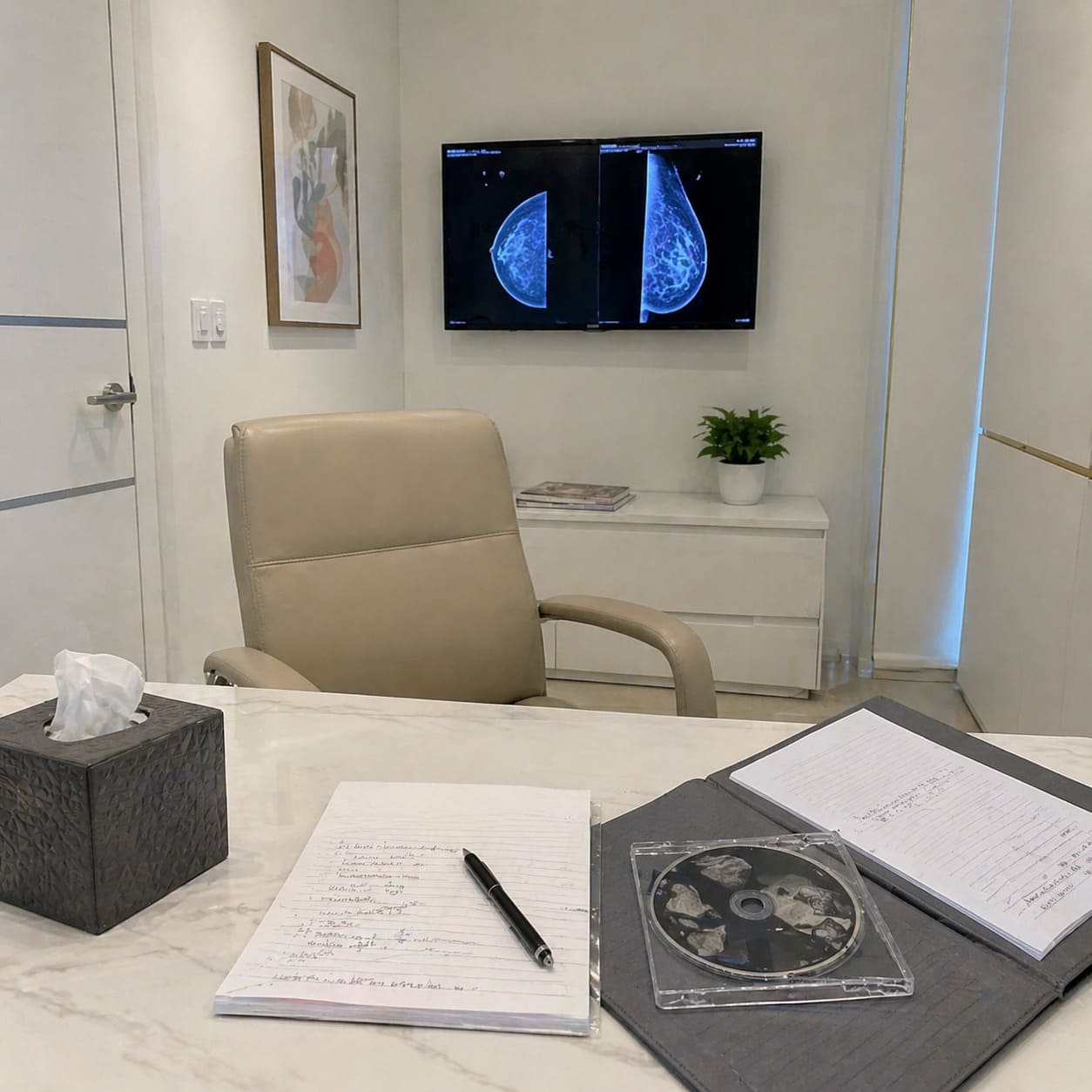
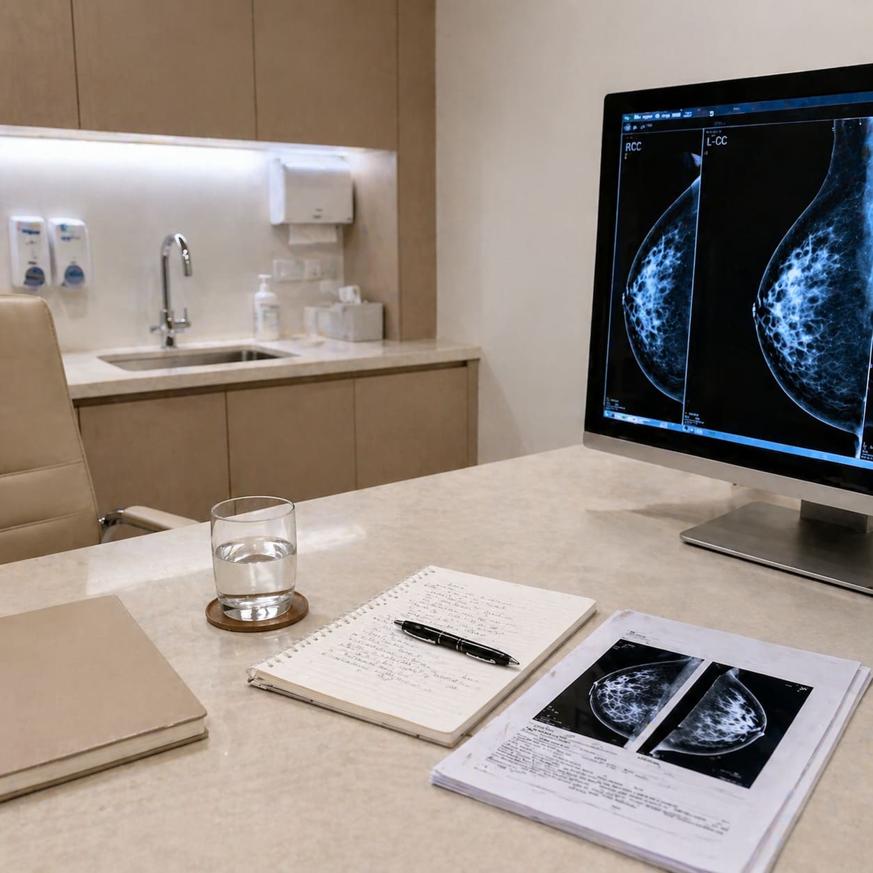
A breast biopsy is done to answer a clear clinical question. Something on an examination or scan may need tissue confirmation because imaging alone cannot always say exactly what a lump, area of distortion, or cluster of calcifications represents.
Sometimes the trigger is a new lump. On other occasions, the concern comes from screening through the NHS Breast Screening Programme, or from a symptom such as nipple change or discharge. Family history and known risk factors may influence how carefully a finding is assessed, but the biopsy itself is usually there to identify what the tissue actually is.
What a biopsy can show
Core needle biopsy is commonly used because it takes a small cylinder of tissue rather than just individual cells. That usually gives more detail about the architecture of the tissue, which helps with lesion characterisation. Fine needle aspiration may be used in selected situations, although it generally gives less structural information.
Radiology and pathology work together here. The scan shows where the abnormal area is and what it looks like. The biopsy shows what it is made of. If those two pieces fit neatly, the diagnosis is more secure.
Even so, a biopsy has limits. The sample is small, and some areas are more mixed than others. A result can answer the main question very clearly, or it can narrow the possibilities and point to further testing. NICE guidance and standard breast clinic practice reflect that same principle: tissue diagnosis is part of the pathway, not the whole pathway.
Expert advice on breast cancer treatment, cosmetic breast surgery, and reconstruction options in London.
Book a ConsultationWhat Happens After an Abnormal Biopsy Result
Once a biopsy result is back, the next stage is usually structured and team-based. Most breast units review the pathology alongside the imaging and clinic findings before making recommendations.
A typical sequence may look like this:
- The pathology report is checked against the scan and examination findings.
- The case may be reviewed at an MDT meeting involving radiology, pathology, surgery, and other relevant specialists.
- The patient is seen or contacted to have the result explained in plain language.
- Further imaging, a repeat biopsy, monitoring, or treatment planning may be advised, depending on what the result shows.
If the report and the imaging tell the same story, the next step is often straightforward. A benign biopsy that matches a clearly benign scan may need reassurance and sometimes follow-up. An indeterminate or atypical finding may lead to more discussion about whether extra sampling or surgery is needed.
Occasionally, the issue is not that the sample is dangerous, but that the pieces do not line up neatly. A pathology result might sound benign, yet the scan still looks suspicious. In that situation, the breast clinic may recommend another biopsy or a different type of tissue sampling because the team is trying to resolve the mismatch, not because anyone is guessing.
Communication matters at this stage. A consultant breast surgeon or another senior clinician will usually explain what was found, how certain the result is, and what the recommendation means in practical terms. In some clinics, a breast care nurse is also part of those discussions, especially if a diagnosis needs more than a short explanation.
For readers seeking a second opinion on a complex or mixed result, services such as D B Ghosh Breast Surgeon Specialist in Cancer and Cosmetic Surgery Harley Street London can be part of that process where appropriate, particularly if the decision is about interpretation, planning, or reconstruction timing rather than speed alone.
Always bring a summary of your key questions to follow-up appointments so nothing important is missed during the discussion.

Types of Abnormal Findings and Their Implications
An abnormal breast biopsy result can sit anywhere on a spectrum from clearly benign to clearly malignant. Management depends on the category, the imaging appearance, and whether the pathology fits the broader clinical picture.
Benign but abnormal findings
Many biopsy results fall into this group. Fibroadenoma, papilloma, fat necrosis, fibrocystic change, and some inflammatory conditions may all be described as abnormal findings because the tissue is not completely typical.
Management varies. A simple fibroadenoma that matches the scan may need no treatment beyond reassurance. A papilloma may prompt further discussion because some are observed and others are removed, depending on associated features and how the diagnosis was made.
Atypia and high-risk changes
Atypical hyperplasia means the cells look unusual and show more change than benign tissue, but they do not meet the criteria for cancer. This matters because atypia can be associated with a higher future risk of breast cancer, and in some situations it may sit close to a more significant area that was not fully captured in the original sample.
Because of that, atypia often leads to a more detailed review. The MDT may consider whether surveillance is appropriate or whether surgical excision would give a clearer answer. Risk stratification is part of that discussion, particularly if family history or other factors are also relevant.
DCIS
Ductal carcinoma in situ, or DCIS, is a non-invasive breast cancer. The cells are malignant, but they remain inside the milk ducts and have not invaded surrounding breast tissue.
That distinction is important. DCIS is cancer in pathological terms, but it does not behave in the same way as invasive cancer. Decisions about surgery and, in some cases, radiotherapy are shaped by the extent of the area, the grade, and where it sits in the breast.
Invasive cancer
An invasive cancer result means cancer cells have moved beyond the ducts or lobules into surrounding breast tissue. The pathology report may describe the type of cancer and may later include other features that guide treatment planning.
At that stage, further tests are often used to build a fuller picture before treatment is finalised. The result may sound stark on the page, but management decisions are usually made in stages, with input from the MDT and support from the breast care team rather than from a single report alone.
If the terminology in your pathology report is unclear, ask your breast care nurse or consultant for a plain language explanation to ensure full understanding.

Common Misconceptions About Abnormal Biopsy Results
A few misunderstandings come up repeatedly after a biopsy result, and they can add unnecessary stress.
- An abnormal result always means cancer. That is one of the commonest misconceptions. Many abnormal breast biopsy results are non-cancerous findings, including benign lesions and tissue changes that simply need correlation with imaging.
- A benign result means nothing else needs checking. Sometimes that is true, but not always. If the biopsy result does not adequately explain what was seen on the scan, the team may still advise more assessment.
- An unclear result means a mistake was made. Breast diagnosis can involve genuine grey areas. Small samples, mixed tissue patterns, and borderline changes can all make interpretation less straightforward. That is why MDT review exists in standard NHS and private breast clinic practice.
- Everything must happen immediately. Some results do require prompt planning, especially confirmed cancer diagnoses. Others need careful review rather than speed. A short period of further discussion or extra imaging does not automatically signal worsening disease.
- A second opinion means starting again from scratch. In reality, a second opinion often involves reviewing the pathology, imaging, and recommendations already made. It is usually about clarity and confidence in the plan, particularly if the findings are atypical, the advice feels mixed, or surgery choices are finely balanced.
Questions to Ask at Your Follow-Up Appointment
A follow-up appointment is often easier if a few key points are written down in advance. The aim is not to cover every possible detail. The aim is to leave with a clear sense of what the result means and what happens next.
- What exactly did the biopsy show in plain language?
- Does the pathology match the scan and examination findings?
- Is the result benign, atypical, in situ, or invasive?
- Do I need any further imaging or another biopsy?
- Will my case be discussed at an MDT meeting, or has it already been reviewed there?
- What are the main options from here, including monitoring if that is relevant?
- If surgery is being considered, what is the reason for it in my case?
- Would a second opinion be reasonable if the result is borderline or the plan is difficult to weigh up?
Some people also find it helpful to ask for a copy of the clinic letter or a summary of the plan. A breast care nurse can often help clarify terms after the main consultation if the conversation feels dense or fast-moving.
Expert breast surgery advice from a leading London consultant with over 30 years of experience.
Book ConsultationLooking Beyond the Result: What an Abnormal Biopsy Really Means for Your Process
An abnormal biopsy result is an important step, but it is rarely the whole story on its own. Breast care decisions are usually made by bringing together the pathology report, radiology, examination findings, and the context of the individual patient.
For some people, the result leads to reassurance and routine follow-up. For others, it opens a more detailed discussion about surgery, surveillance, or risk reduction. The same word, abnormal, can therefore cover very different situations.
Context changes meaning. A benign lesion that fits perfectly with the scan may close the investigation neatly. An atypical result may call for more tissue to be examined. A cancer diagnosis may still involve several decisions about type of surgery, timing, and whether reconstruction is relevant.
That is one reason consultant-led review matters in complex breast care. In settings such as D B Ghosh Breast Surgeon Specialist in Cancer and Cosmetic Surgery Harley Street London, the value often lies in careful interpretation and planning, especially where the wording of the report feels more confusing than helpful.
Seen in that wider frame, an abnormal biopsy is best understood as a signpost in a managed clinical pathway. It points to the next decision, not to a fixed outcome, and the most useful next step is a clear explanation of how your result fits the bigger picture of your breast health.