
Do calcifications on your mammogram mean breast cancer?
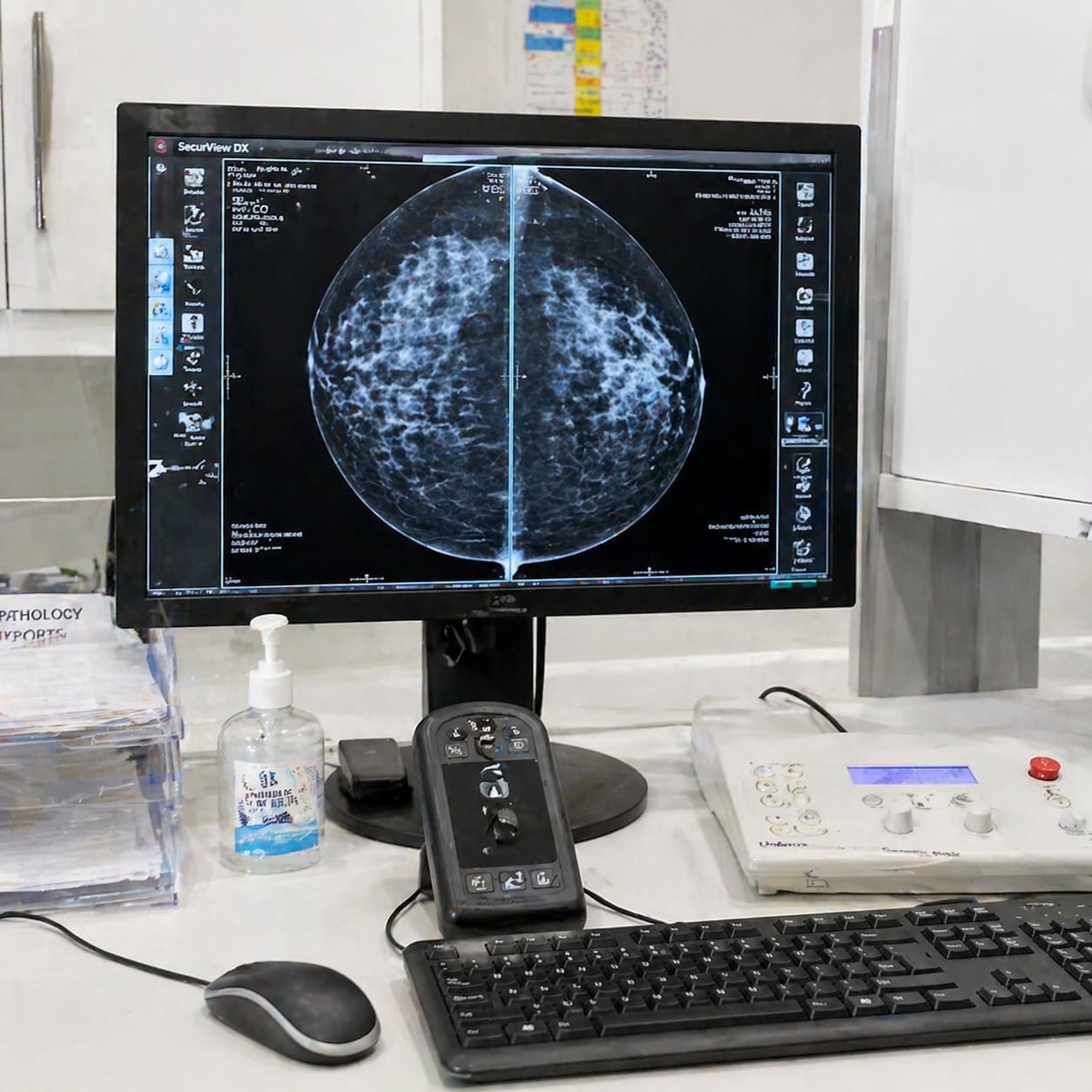
Calcifications on a mammogram do not automatically mean breast cancer. They are tiny calcium deposits, usually seen only on imaging, and the next step depends on their size, shape, pattern and whether they sit in a cluster. We treat a recall as a need for clearer assessment, not a diagnosis.
The short answer is usually no
You may have opened a mammogram report and seen the words “calcifications” or “microcalcifications” without much explanation. We know those words can sound heavier than they are. Calcifications are an imaging finding, which means they describe something seen on the mammogram rather than naming a disease.
Breast calcifications are small calcium deposits in the breast. Macmillan Cancer Support explains that they do not cause symptoms and cannot be felt as a lump, which is why they are usually picked up on a routine mammogram or during breast screening.
A breast screening recall for calcifications means the radiologist wants a clearer look at the area. Sometimes that ends with reassurance. Sometimes it leads to more mammogram views or a biopsy. The reason lies in the appearance of the calcifications, so the useful question is what they look like and how they are arranged.
Expert advice on breast cancer treatment, cosmetic breast surgery, and reconstruction options in London.
Book a ConsultationMacrocalcifications and microcalcifications mean different things
Two main types of breast calcification appear on mammograms, and they do not carry the same meaning. Macrocalcifications are usually benign-looking and do not need treatment or monitoring. Microcalcifications are smaller, and their pattern needs closer reading.
| Macrocalcifications | Microcalcifications |
|---|---|
| Usually larger calcium deposits seen on mammogram. | Smaller calcium deposits that need closer interpretation. |
| Usually harmless and not linked to cancer. | Most are benign, but a cluster in one area may signal pre-cancerous change or early breast cancer. |
| More common after the menopause and can develop naturally with age. | Their shape, grouping and spread guide whether further tests are sensible. |
| Usually need no treatment or monitoring when they have benign features. | May lead to magnification views or biopsy if the radiologist cannot classify them confidently. |
Words such as amorphous, pleomorphic or linear on a report are descriptors of shape and pattern. They do not give you the full answer on their own. A radiologist reads those words alongside the images, previous mammograms where available, and any related breast change.
At D B Ghosh, we spend time translating unclear mammogram wording into the decision it points to, because “clustered microcalcifications” is not the same thing as a cancer diagnosis. The wording matters, but the image pattern matters more.

If a report mentions clustered microcalcifications, compare it with any previous mammograms before deciding how significant the change is. Stability over time can affect the level of concern.

The pattern decides the level of concern
Calcium in the breast is common enough that we do not judge calcifications by their presence alone. We look at how they behave on imaging, including their size, their shape, their pattern, and whether they form a group that follows a concerning line or area.
A radiologist assessing mammogram calcifications is asking a practical question. Do these deposits look like a benign ageing change, or do they have features that need proof from a tissue sample? NHS England breast screening assessment guidance supports further mammogram views for calcifications, including lateral and magnification views, because those views help define their extent and appearance more clearly.
Comparison with older images can change the reading. Calcifications that have looked the same over time may be treated differently from new calcifications or calcifications that have changed. A related mass, area of distortion or tissue change also alters the discussion, because the calcifications no longer sit as an isolated finding.
Newer imaging has a place in breast assessment, but we keep this point precise. For microcalcifications, traditional supplementary mammographic views remain the standard of care in NHS England guidance. Digital breast tomosynthesis can be useful in assessment of some screen-detected soft tissue abnormalities, but it has not replaced magnification views for microcalcifications.
London-based consultant breast and oncoplastic care is useful when the question has moved from “what is this word?” to “does this report, the imaging and the recommended next step make sense together?” Suspicious means the finding needs evidence. It does not mean the answer has already been decided.

Biopsy is used to prove what the mammogram suggests
When the image remains uncertain after assessment, a biopsy may be advised to test the tissue that corresponds to the calcifications. That step is used when imaging cannot safely place the finding in the benign category.
A stereotactic biopsy, also called a mammogram-guided biopsy, uses mammogram targeting to sample calcifications that may not show well on ultrasound. A core biopsy removes small tissue samples for pathology, which means laboratory examination by a pathologist. Vacuum-assisted biopsy is another mammogram-guided method used in some situations to obtain a larger sample.
The sequence matters because the sample has to explain the image:
- We start with the imaging concern, including where the calcifications sit and how far they appear to extend.
- Targeted sampling is planned so the biopsy reaches the area seen on the mammogram.
- Specimen radiography, which is an X-ray of the biopsy sample, checks that representative calcification is present in the tissue taken.
- A pathologist examines the tissue and reports whether the finding is benign, uncertain, pre-cancerous or cancerous.
- Imaging and pathology are then matched. If they do not fit, further sampling may be needed.
NHS England breast screening assessment guidance describes triple assessment as imaging, clinical examination and image-guided needle biopsy for tissue diagnosis where indicated. It also says each patient who has had a needle biopsy should be discussed at a multidisciplinary team meeting, sometimes shortened to MDT, where the imaging, clinical findings and pathology are correlated.
At D B Ghosh, we place that matching step at the centre of planning. A biopsy result is strongest when it answers the original mammogram question, because surgery or follow-up should be built on evidence that fits.
A benign biopsy is only reassuring when it matches the mammogram pattern. If the pathology and imaging do not fit together, correlation matters as much as the biopsy label.

A result is useful only when it explains the finding
A benign biopsy result is reassuring when it matches the mammogram appearance. If the result says the tissue is harmless but the imaging still looks concerning, the right response is not to ignore the mismatch. We want the radiologist, pathologist and breast surgeon to agree that the sample explains the finding.
If cancer cells are found after biopsy of microcalcifications, the diagnosis is usually ductal carcinoma in situ, called DCIS, or a very small early breast cancer. DCIS means a non-invasive breast cancer, where the abnormal cells are inside the breast ducts. Treatment planning then depends on the extent of calcifications, the biopsy result, breast size and shape, and whether breast conservation is a safe option.
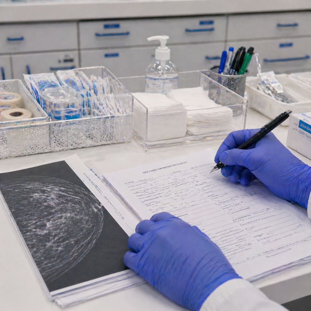
A second opinion is sensible when the report wording is unclear, calcifications are extensive, biopsy results and imaging seem out of step, or surgery has been recommended before you feel the plan has been properly explained. In clinic, useful questions include whether the calcifications look benign or suspicious, whether previous mammograms have been compared, whether the biopsy sampled the right area, and how the result changes the surgical plan if DCIS or early cancer is found.
Speed has value, but it is not the main standard. The priority is correlation: the report, images, biopsy and plan should tell one coherent story before treatment decisions are made.
Expert breast surgery advice from a leading London consultant with over 30 years of experience.
Book ConsultationCommon questions about breast calcifications
Can calcium tablets or dairy cause breast calcifications?
No. Calcium in your diet does not cause breast calcifications on a mammogram, so you should not assume supplements or high-calcium foods are the reason for the finding.
Do macrocalcifications need follow-up?
Macrocalcifications with benign features are usually harmless and do not need treatment or monitoring. We still read them in the context of the whole mammogram, but they are different from microcalcifications that need closer assessment.
Are clustered microcalcifications always cancer?
No. Most microcalcifications are benign, but a cluster in one area may be linked with pre-cancerous change or early breast cancer. That is why the pattern is assessed carefully before deciding on biopsy or follow-up.
Can a normal ultrasound rule out suspicious microcalcifications?
No. Suspicious microcalcifications are mammogram findings, and they may not show clearly on ultrasound. A normal ultrasound does not automatically cancel out a mammogram concern.
Why might a biopsy marker or clip be placed after sampling?
A small marker can be placed to show exactly where the biopsy was taken. That helps the team match the biopsy result back to the mammogram finding and plan any further care accurately.
This is general information, not medical advice.